42 label eye diagram
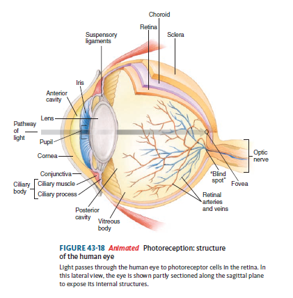
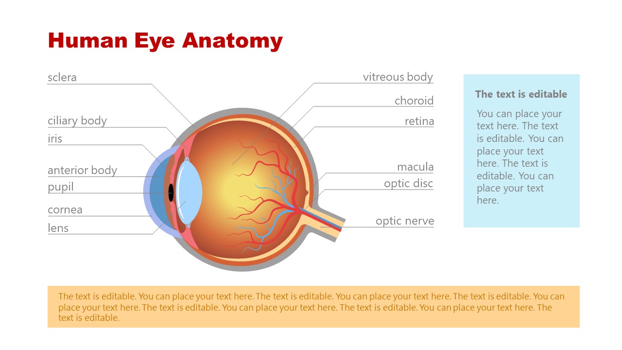
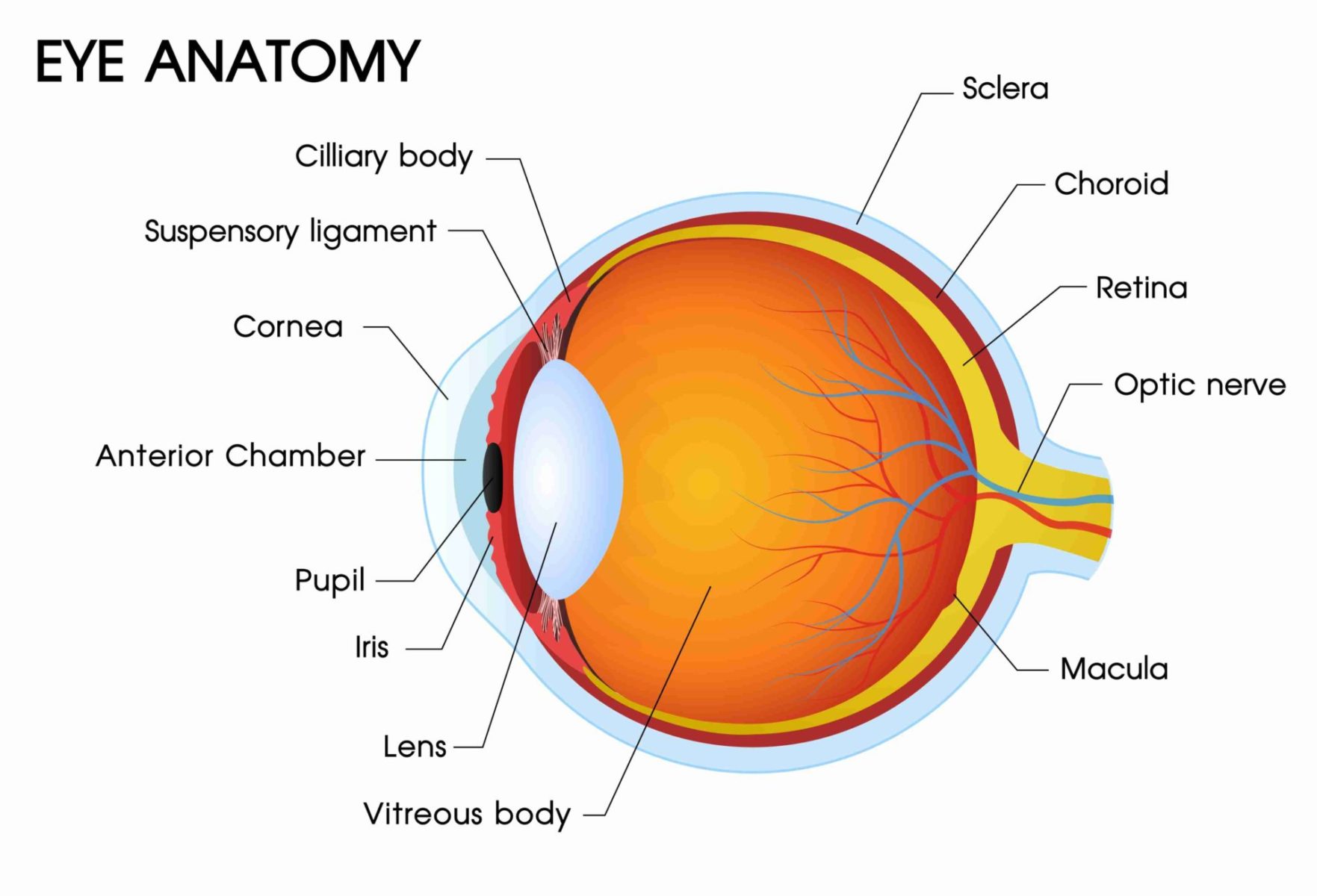
Label the Eye Diagram | Quizlet Start studying Label the Eye. Learn vocabulary, terms, and more with flashcards, games, and other study tools. Labelled Diagram of Human Eye, Explanation and Function - Vedantu Labeled Diagram of Human Eye The eyes of all mammals consist of a non-image-forming photosensitive ganglion within the retina which receives light, adjusts the dimensions of the pupil, regulates the availability of melatonin hormones, and also entertains the body clock.
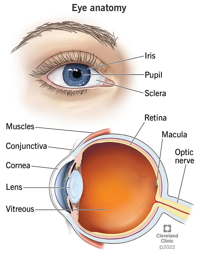
Eye Anatomy: Parts of the Eye and How We See Eye Anatomy: Parts of the Eye Outside the Eyeball The eye sits in a protective bony socket called the orbit. Six extraocular muscles in the orbit are attached to the eye. These muscles move the eye up and down, side to side, and rotate the eye. The extraocular muscles are attached to the white part of the eye called the sclera.
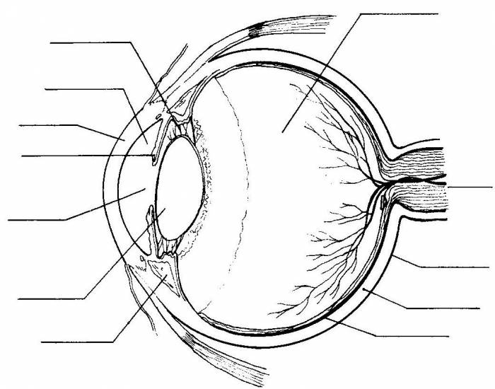
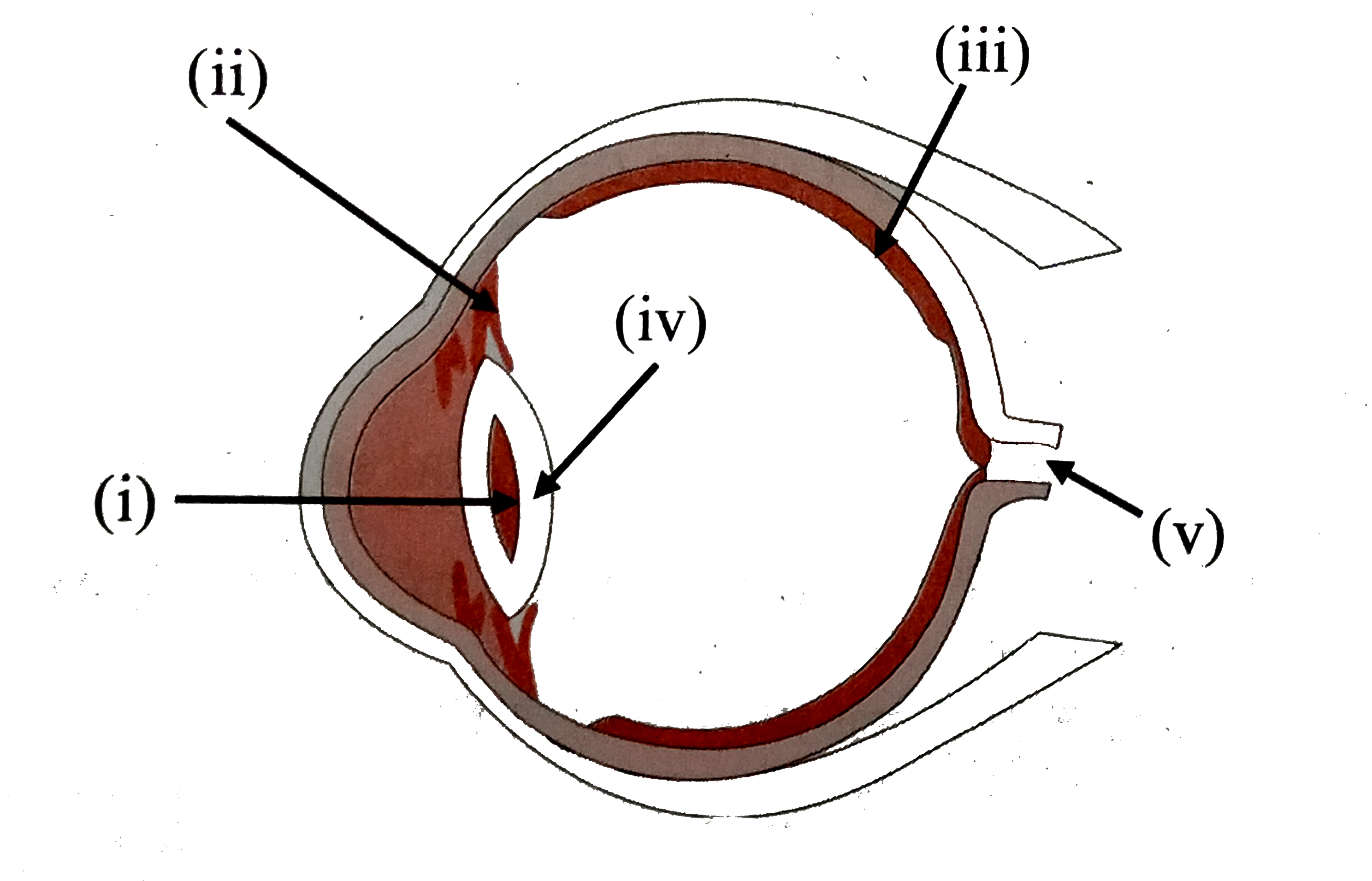
Label eye diagram
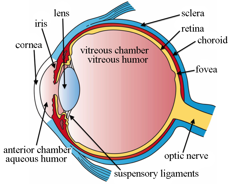
Label Parts of the Human Eye - University of Dayton Label Parts of the Human Eye Parts of the Eye Select the correct label for each part of the eye. The image is taken from above the left eye. Click on the Score button to see how you did. Incorrect answers will be marked in red. 280+ Labeled Eye Stock Photos, Pictures & Royalty-Free Images - iStock Browse 280+ labeled eye stock photos and images available, or start a new search to explore more stock photos and images. Sort by: Most popular. Eye anatomy with labeled structure scheme for human optic... Eye anatomy with labeled structure scheme for human optic outline diagram. Educational physiological and medical sight infographic with side ... eye labeling Diagram | Quizlet fibrous transparent layer of clear tissue like a dome that covers the anterior portion of the eyeball (the iris and pupil). It is the first structure to refract (bend) light that enters the eye. Location Term sclera Definition Tough white out covering of the eyeball Location Term choroid Definition
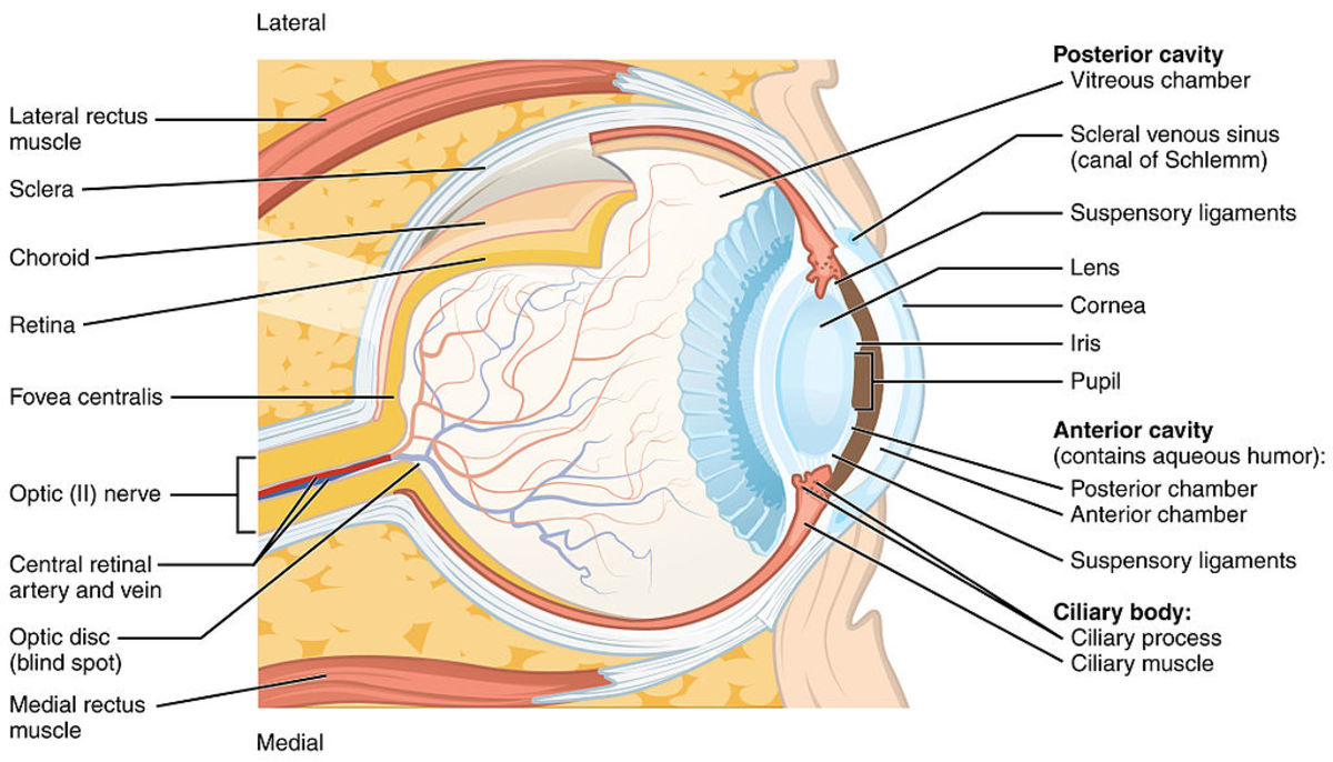
Label eye diagram. Simple eye diagrams | Easy eye diagram | Labeled eye ... - Pinterest Also labeled eye diagram and anatomy of eye and human eye structure for better understanding. Human eye diagram and functions with diagram of human eye with ... What Does the Eye Look Like? - Diagram of the Eye | Harvard Eye Associates Anterior Chamber: The space between the cornea and the iris, filled with the aqueous humor. Choroid: The vascular layer of the eye, containing connective tissue. Nutrition of the eye is dependent upon blood vessels in the choroid. Ciliary Body: the part of the eye that connects the iris to the choroid. Label the Eye Quiz - PurposeGames.com Label the Eye by LegoA1 393,263 plays 12 questions ~30 sec English 12p More 163 4.21 (you: not rated) Tries Unlimited [?] Last Played April 12, 2023 - 08:25 PM There is a printable worksheet available for download here so you can take the quiz with pen and paper. From the quiz author Title Says It ALL!!!! Remaining 0 Correct 0 Wrong 0 Press play! Labelling the eye — Science Learning Hub Use this interactive to label different parts of the human eye. Drag and drop the text labels onto the boxes next to the diagram. Selecting or hovering over a box will highlight each area in the diagram. Cornea Vitrous humour Lens Pupil Retina Iris Schlera Optic nerve Download Exercise
Anatomy of the eye: Quizzes and diagrams | Kenhub Take a look at the diagram of the eyeball above. Here you can see all of the main structures in this area. Spend some time reviewing the name and location of each one, then try to label the eye yourself - without peeking! - using the eye diagram (blank) below. Unlabeled diagram of the eye 160+ Labeled Eye Illustrations, Royalty-Free Vector Graphics ... - iStock Browse 160+ labeled eye stock illustrations and vector graphics available royalty-free, or start a new search to explore more great stock images and vector art. Sort by: Most popular. Eye anatomy with labeled structure scheme for human optic... Eye anatomy with labeled structure scheme for human optic outline diagram. Structure and Functions of Human Eye with labelled Diagram - BYJU'S Human Eye Diagram: Contrary to popular belief, the eyes are not perfectly spherical; instead, it is made up of two separate segments fused together. Explore: Facts About The Eye To understand more in detail about our eye and how our eye functions, we need to look into the structure of the human eye. Recommended Video: 1,221 Cow's Eye Dissection | Exploratorium The lens in your eye bends the light that has reflected from that tree to make a perfect little upside-down picture of the tree on the back of your eyeball. ... Instructions include an eye diagram, a glossary, and color photos for each step. File. coweye.pdf (326.92 KB) Credits & Use Policy . Producer: Noah Wittman.
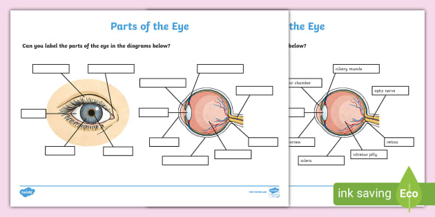
PDF National Eye Institute | National Eye Institute National Eye Institute | National Eye Institute Eye Diagram With Labels and detailed description - BYJU'S Diagram Of Eye Diagram Of Eye The human eye is responsible for the most important function of the human body, the sense of sight. It consists of several distinct parts that work in coordination with each other. The most common eye diseases include myopia, hypermetropia, glaucoma and cataract. The Eyes (Human Anatomy): Diagram, Optic Nerve, Iris, Cornea ... - WebMD The Eyes (Human Anatomy): Diagram, Optic Nerve, Iris, Cornea, Pupil, & More Menu Eye Health Reference A Picture of the Eye Written by WebMD Editorial Contributors Medically Reviewed by... Label the Eye Worksheet - Teacher-Made Learning Resources - Twinkl The first page is a labelling exercise with two diagrams of the human eye. One is a view from the outside, and the other is a more detailed cross-section. Challenge learners to label the parts of the eye diagram. Show more Related Searches eye parts of the eye part of the eye human eye diagram eye diagram labelling the eye Ratings & Reviews
PDF Parts of the Eye - National Institutes of Health Eye Diagram Handout Author: National Eye Health Education Program of the National Eye Institute, National Institutes of Health Subject: Handout illustrating parts of the eye Keywords: parts of the eye, eye diagram, vitreous gel, iris, cornea, pupil, lens, optic nerve, macula, retina Created Date: 12/16/2011 12:39:09 PM
Human Eye Anatomy Pictures, Images and Stock Photos - iStock Parts of the eye, labeled vector illustration diagram. Educational beauty and nursing information. Eyelid, eyelashes, pupil, lacrimal gland and other ...
FREE! - The Human Eye Labeling Activity (Teacher-Made) - Twinkl In this resource, you'll find a 2-page PDF that is easy to download, print out, and use immediately with your class. The first page is a labelling exercise with two diagrams of the human eye. One is a view from the outside, and the other is a more detailed cross-section. Challenge learners to label the parts of the eye diagram. Show more.
Anatomy of the Human Eye - News Medical Eyes are one of the most important organs of the body. A healthy pair of eyes means a clear vision, which plays a major role in day-to-day life and quality ...
Label Eye Printout - EnchantedLearning.com Label the Eye Diagram. Human Anatomy. Read the definitions, then label the eye anatomy diagram below. Cornea - the clear, dome-shaped tissue covering the front of the eye. Iris - the colored part of the eye - it controls the amount of light that enters the eye by changing the size of the pupil. Lens - a crystalline structure located just behind ...
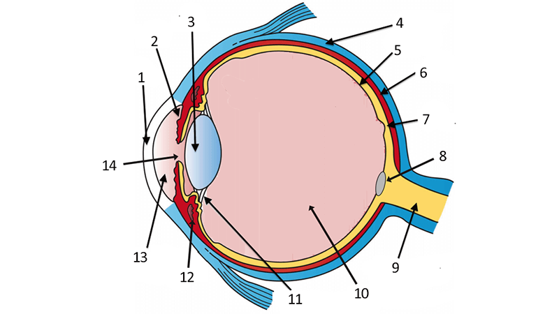
Label the Eye - The Biology Corner The image was modified from an eye diagram at Wikimedia Commons. I added the numbers and additional errors to identify structures that weren't on the original diagram. There are a few terms that can be vague, for example, the aqueous humor could also be labeled as the aqueous chamber. Zonule of Zinn can also be called suspensory ligaments.
Eye Anatomy Labeling royalty-free images - Shutterstock Eye anatomy with labeled structure scheme for human optic outline diagram. · Parts of the eye, labeled vector illustration diagram. · Vintage anatomy posters.
Eye Diagram Pictures, Images and Stock Photos - iStock Parts of the eye, labeled vector illustration diagram. Educational beauty and nursing information. Eyelid, eyelashes, pupil, lacrimal gland and other ...
The Eye - Science Quiz - Seterra - GeoGuessr The anatomy of the eye is fascinating, and this quiz game will help you ... Light enters our eyes through the pupil, then passes through a lens and the ...
eye labeling Diagram | Quizlet fibrous transparent layer of clear tissue like a dome that covers the anterior portion of the eyeball (the iris and pupil). It is the first structure to refract (bend) light that enters the eye. Location Term sclera Definition Tough white out covering of the eyeball Location Term choroid Definition
280+ Labeled Eye Stock Photos, Pictures & Royalty-Free Images - iStock Browse 280+ labeled eye stock photos and images available, or start a new search to explore more stock photos and images. Sort by: Most popular. Eye anatomy with labeled structure scheme for human optic... Eye anatomy with labeled structure scheme for human optic outline diagram. Educational physiological and medical sight infographic with side ...
Label Parts of the Human Eye - University of Dayton Label Parts of the Human Eye Parts of the Eye Select the correct label for each part of the eye. The image is taken from above the left eye. Click on the Score button to see how you did. Incorrect answers will be marked in red.
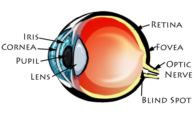
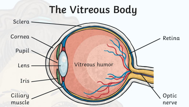


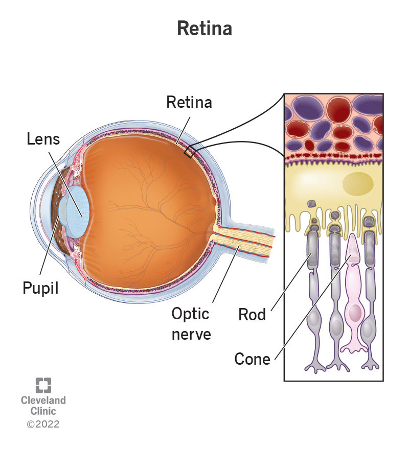











Komentar
Posting Komentar